

Optical stent inspection of surface texture and coating thickness

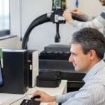
Carlos has collaborated since 2010 on the development of confocal, interferometry and focus variation technologies at Sensofar, where he holds the R&D Engineering Manager position since 2018. His interests are Optomechanical Systems Design and Image Processing.
Its consolidated research work in optical engineering confer the Sensofar R&D group an outstanding position to always stay up-to-date in terms of innovation and the highest technological level.


Optical stent inspection of surface texture and coating thickness full article
C. Bermudez,1,2 F. Laguarta,1 C. Cadevall,1,2 A. Matilla,2 S. Ibañez,3 R. Artigas1,2
1 Universitat Politècnica de Catalunya (UPC) Rambla Sant Nebridi, 10, E-08222 Terrassa, Spain
2 Sensofar Tech SL, (Spain)
3 Sensofar Medical SL, (Spain)
Proc. SPIE 10110, Photonic Instrumentation Engineering IV, 1011007 (20 February 2017)
Abstract
Stent quality control is a critical process. Coronary stents have to be inspected 100% so no defective stent is implanted into a human body. We have developed a high numerical aperture optical stent inspection system able to acquire both 2D and 3D images. Combining a rotational stage, an area camera with line-scan capability and a triple illumination arrangement, unrolled sections of the outer, inner, and sidewalls surfaces are obtained with high resolution. During stent inspection, surface roughness and coating thickness uniformity is of high interest. Due to the non-planar shape of the surface of the stents, the thickness values of the coating need to be corrected with the 3D surface local slopes.
A theoretical model and a simulation are proposed, and a measurement with white light interferometry is shown. Confocal and spectroscopic reflectometry showed to be limited in this application due to stent surface roughness. Due to the high numerical aperture of the optical system, only certain parts of the stent are in focus, which is a problem for defect detection, specifically on the sidewalls. In order to obtain fully focused 2D images, an extended depth of field algorithm has been implemented.
A comparison between pixel variance and Laplacian filtering is shown. To recover the stack image, two different methods are proposed: maximum projection and weighted intensity. Finally, we also discuss the implementation of the processing algorithms in both the CPU and GPU, targeting real-time 2-Million pixel image acquisition at 50 frames per second.
References
Carlos Bermudez, Ferran Laguarta, Cristina Cadevall, Aitor Matilla, Sergi Ibañez, and Roger Artigas. Stent optical inspection system calibration and performance, 2017, Applied Optics 56(9) D134-D141
Carlos Bermudez, Ferran Laguarta, Cristina Cadevall, Aitor Matilla, Sergi Ibañez and Roger Artigas. Novel Stent Optical Inspection System, 2016, Applied Industrial Optics: Spectroscopy, Imaging and Metrology, Paper# AITh2B.3


