Optical profilometry in forensic taphonomy: studying cannibalism and interpersonal violence in archaeological contexts
Francesc Marginedas, Miguel Ángel Moreno-Ibáñez, Antonio Rodríguez-Hidalgo and Palmira Saladié
The Catalan Institute of Human Paleoecology and Social Evolution (IPHES) is an institution that promotes transdisciplinary and advanced research, education, and knowledge transfer. His fields combine the humanities, social sciences, geosciences, and biosciences to study human and social evolution.
The integration of Sensofar 3D optical profilometry into forensic taphonomy research provides powerful tools for the quantitative study of bone surface modifications.
Forensic taphonomy studies the processes that affect human remains from the moment of death to their discovery. Within this research line at IPHES-CERCA, advanced microscopy and 3D surface analysis are used to understand bone-surface modifications related to human behavior, violence, cannibalism, and funerary practices in archaeological contexts.
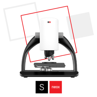
A central methodological tool in this research is the Sensofar S neox 3D optical profilometer, which enables high-resolution three-dimensional reconstruction and quantitative analysis of bone surface modifications.
The integration of optical profilometry with archaeological and forensic approaches allows researchers to distinguish anthropogenic marks from natural taphonomic processes, providing objective evidence for interpreting past human behavior.
Our forensic taphonomy research integrates osteological analysis, microscopic observation, and quantitative 3D surface measurements. Bone surface modifications are first identified macroscopically and with stereomicroscopy. Selected specimens are then analyzed using Confocal microscopy and optical profilometry for detailed 3D reconstructions of the marks.
The Sensofar S neox optical profilometer is used to generate high‑resolution topographic models of bone surfaces. These models allow precise measurement of cut‑mark morphology, including width, depth, cross‑sectional geometry, wall angles, and surface microtopography. Quantitative measurements can then be compared with experimental reference collections and statistically analyzed to determine the origin of the modifications.
MEASUREMENTS
Cultural cannibalism at Maszycka Cave (Poland)
The Maszycka Cave assemblage, dated to approximately 18,000 years ago and attributed to Magdalenian hunter‑gatherers, contains numerous human skeletal remains that show evidence of anthropogenic modification. The study focused on identifying whether bone surface marks corresponded to butchery activities associated with cannibalism.

Using the Sensofar S neox optical profilometer, high‑resolution scans of bone surface modifications were obtained (Figure 1 a, b). Cross‑sections of the cut marks were extracted and analyzed morphometrically, measuring parameters such as incision width, depth, wall length, and cross‑sectional angles (Figure 1 c, d).

These quantitative data were compared with experimental marks produced by stone tools, carnivore tooth marks, and trampling marks. Multivariate statistical analyses demonstrated that the archaeological marks closely matched experimentally produced cut marks, confirming their anthropogenic origin (Figure 2). The results showed that the human remains were intensively processed shortly after death for the extraction of meat, brain tissue, and marrow, providing strong evidence for cannibalistic practices among Magdalenian groups.

Interpersonal violence at Roc de les Orenetes (Pyrenees, Spain)
Another research application involves the analysis of perimortem trauma in the collective burial site of Roc de les Orenetes, located in the Eastern Pyrenees at an altitude of 1840 meters. The assemblage contains thousands of disarticulated human bones representing at least 51 individuals, dating to the Late Chalcolithic and Early Bronze Age. The study aimed to determine whether observed bone lesions resulted from interpersonal violence rather than funerary manipulation or post-depositional processes. Bone trauma was analyzed using stereomicroscopy, digital microscopy, and the Sensofar S neox Confocal optical profilometer.

Three-dimensional surface analysis enabled detailed characterization of sharp-force trauma, including chop and slice marks produced by bladed weapons or projectiles. The optical profilometer enabled precise visualization of incision geometry and penetration depth, supporting the identification of weapon-related injuries and differentiating them from other types of bone modification (Figure 3).

The results revealed numerous perimortem injuries concentrated on ribs and upper limbs, suggesting episodes of interpersonal violence involving bladed weapons and projectile points. The combination of taphonomic analysis and high-resolution surface imaging allowed a detailed reconstruction of the violent events affecting this prehistoric population.
Technological contribution of Sensofar optical profilometry
The use of Sensofar optical profilometry has significantly advanced the study of bone surface modifications in archaeological and forensic contexts.
The technology provides:
- Non‑destructive high‑resolution 3D surface measurements.
- Accurate reconstruction of cut‑mark cross‑sections.
- Quantitative morphometric data for statistical analysis.
- Improved discrimination between anthropogenic and natural modifications.
- Detailed visualization of trauma produced by different types of tools or weapons.
By integrating optical profilometry into forensic taphonomy workflows, researchers can achieve a more robust and objective interpretation of bone surface modifications.
The integration of Sensofar 3D optical profilometry into forensic taphonomy research provides powerful tools for the quantitative study of bone surface modifications. Applications at sites such as Maszycka Cave and Roc de les Orenetes demonstrate how high-resolution surface analysis can clarify complex archaeological interpretations of cannibalism, violence, and past human behavior.
This research line highlights the importance of advanced surface metrology technologies in archaeology and forensic anthropology, enabling researchers to investigate ancient human activities with unprecedented precision.
References
[1] Marginedas, F., Saladié, P., Połtowicz-Bobak, M., Terberger, T., Bobak, D. and Rodríguez-Hidalgo, A. (2025). New insights of cultural cannibalism amongst Magdalenian groups at Maszycka Cave, Poland. Scientific Reports, 15: 2351.
[2] Moreno-Ibáñez, M.Á., Saladié, P., Ramírez-Pedraza, I., Díez-Canseco, C., Fernández-Marchena, J.L., Soriano, E., Carbonell, E. and Tornero, C. (2024). Death in the high mountains: Evidence of interpersonal violence during Late Chalcolithic and Early Bronze Age at Roc de les Orenetes (Eastern Pyrenees, Spain). American Journal of Biological Anthropology, 184 (1): e24909.
Related products